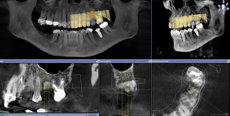
Dental Cone Beam CT
Dental cone beam computed tomography (CT) is a special type of x-ray equipment used when regular dental or facial x-rays are not sufficient. Your doctor may use this technology to produce three dimensional (3-D) images of your teeth, soft tissues, nerve pathways and bone in a single scan.

This procedure requires little to no special preparation. Tell your doctor if there's a possibility you are pregnant. Wear loose, comfortable clothing and leave jewelry at home. You may be asked to wear a gown.
01. What is Dental Cone Beam CT?
Dental cone beam computed tomography (CT) is a special type of x-ray machine used in situations where regular dental or facial x-rays are not sufficient. It is not used routinely because the radiation exposure from this scanner is significantly more than regular dental x-rays. See the Safety page for more information about x-rays. This type of CT scanner uses a special type of technology to generate three dimensional (3-D) images of dental structures, soft tissues, nerve paths and bone in the craniofacial region in a single scan. Images obtained with cone beam CT allow for more precise treatment planning.
02. What are some common uses of the procedure?
- Surgical planning for impacted teeth.
- Diagnosing temporomandibular joint disorder (TMJ).
- Accurate placement of dental implants.
- Evaluation of the jaw, sinuses, nerve canals and nasal cavity.
- Detecting, measuring and treating jaw tumors.
- Determining bone structure and tooth orientation.
- Locating the origin of pain or pathology.
- Cephalometric analysis.
- Reconstructive surgery.
03. How does the procedure work?
During a cone beam CT examination, the C-arm or gantry rotates around the head in a complete 360-degree rotation while capturing multiple images from different angles that are reconstructed to create a single 3-D image.
The x-ray source and detector are mounted on opposite sides of the revolving C-arm or gantry and rotate in unison. In a single rotation, the detector can generate anywhere between 150 to 200 high resolution two-dimensional (2-D) images, which are then digitally combined to form a 3-D image that can provide your dentist or oral surgeon with valuable information about your oral and craniofacial health.
04. What will I experience during and after the procedure?
You will not experience any pain during a cone beam CT exam, and you will be able to return to your normal activities once the exam is complete.


